3D MEDICAL IMAGING SOFTWARE
3D Medical Imaging Software Development
Our team built a 3D medical imaging software for reconstruction of bones, skin, and various organs from X-rays and CT scans implementing machine learning.
#Healthcare
#IoT
#ComputerVision
Client*
A healthcare technology firm producing advanced devices and software that support healthcare professionals in their everyday tasks.
*We cannot provide any information about the client or specifics of the case study due to non-disclosure agreement (NDA) restrictions.
Project in numbers
duration
Ongoing project
12 specialists
Team involved in the project
industry
Healthcare
solution
Web
Python, FastAPI, PyQt, JavaScript, React, MS SQL Server, Weights and Biases, MLFlow, PyTorch, OpenCV, TensorFlow, Keras, ONNXRuntime, PyDICOM, Albumentations, AWS (S3, EC2, Lambda), AWS SageMaker (Studio, Model Monitoring, Inference endpoint), Qase, Postman, Swagger, TestFlight, Arduino, Thonny
Services
2 x Front-end developers
2 x Back-end developers
1 x Project manager
4 x ML engineers
2 x QA specialists
1 x UX/UI Designer
Challenge
Develop an ML-based tool for 3D medical imaging of bones, skin, and other body parts by converting flat scans into three-dimensional volumetric models from X-rays and CT scans. By transforming flat scans into detailed 3D volumetric models, healthcare professionals could gain enhanced visibility for patient treatment and deeper insights into diseases and abnormalities.
Solution & functionality
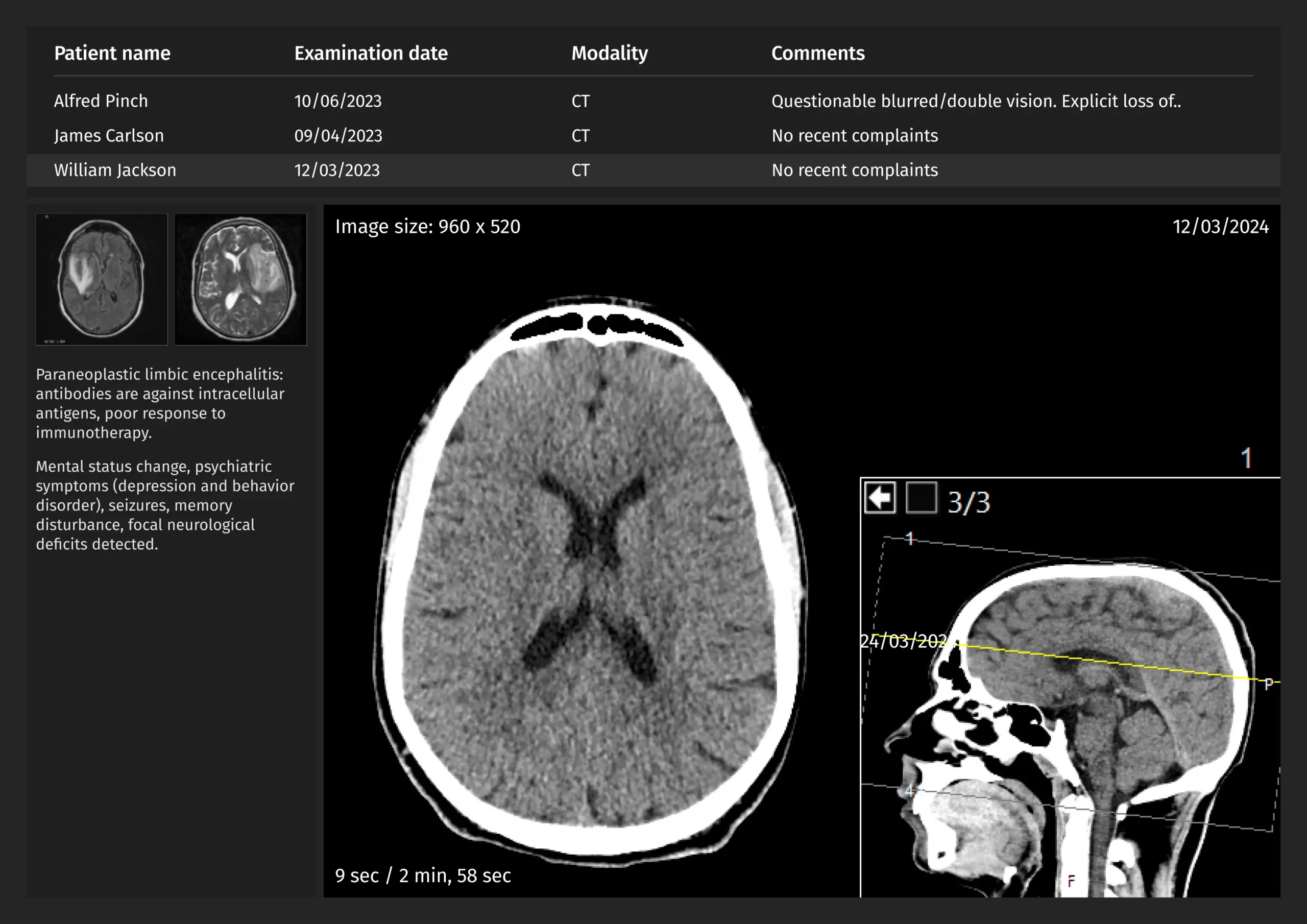
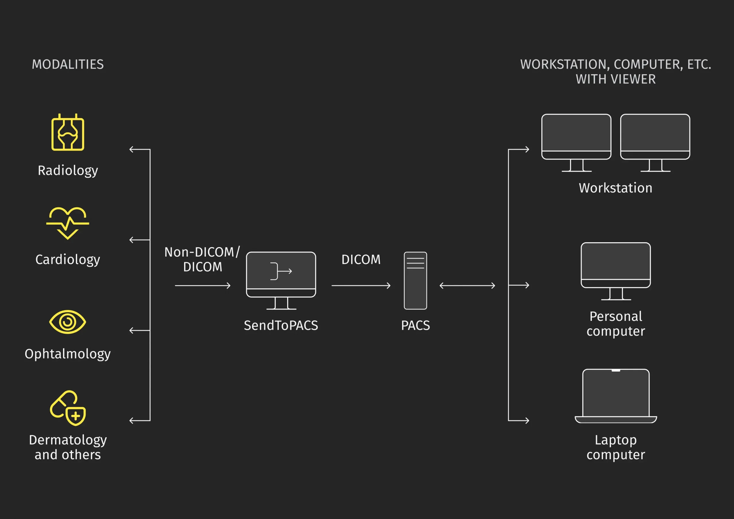
We integrated medical imaging analysis into the customer’s system, ensuring compatibility with X-rays and CT scans from radiology, cardiology, and other labs. As a result, all the 3D medical images can be accessed across hospital workstations and personal laptops.
3D rendering for X-rays and CT scans
Conversion of black-and-white images into 3D medical models takes just a few clicks. Once the X-ray or CT scan is uploaded, clinicians can set threshold attenuation values to define 3D detail, let the platform scan each piece, and create voxels reconstructing denser body fragments. This results in volumetric 3D medical images.
After rendering, clinicians can use a toolbar to manage objects: zoom in/out, add/remove skin, tissue, muscles, and bones, and cut away excess parts. The primary tool, a cube, allows the image rotation for a more accurate view of the pathology.
Compatibility and security for DICOM files
Initially, we ensured that the web platform effortlessly worked with DICOM files, the standard format for medical imaging management. Next, we enhanced security to safeguard the confidential health information they carry.
Our developers built a secure space to store imported DICOM files, encompassing patient details, diagnoses, treatments, dates, and test results.
ROI manager for highlighting pathologies
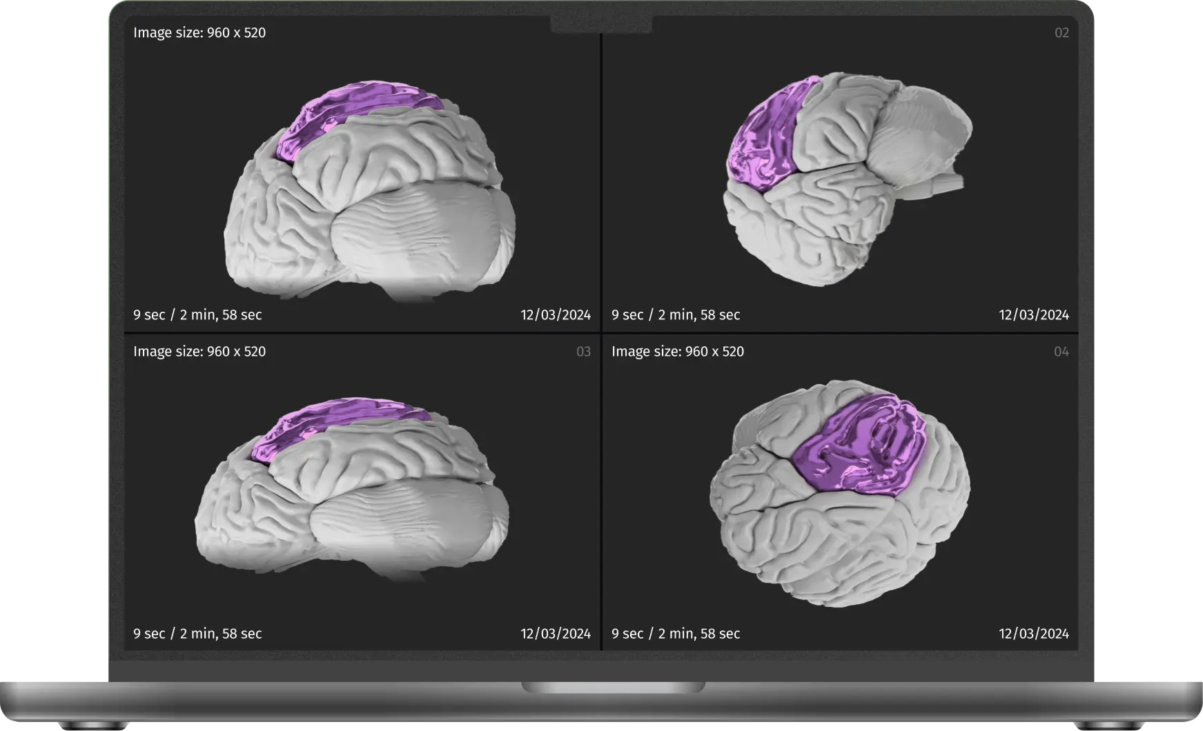
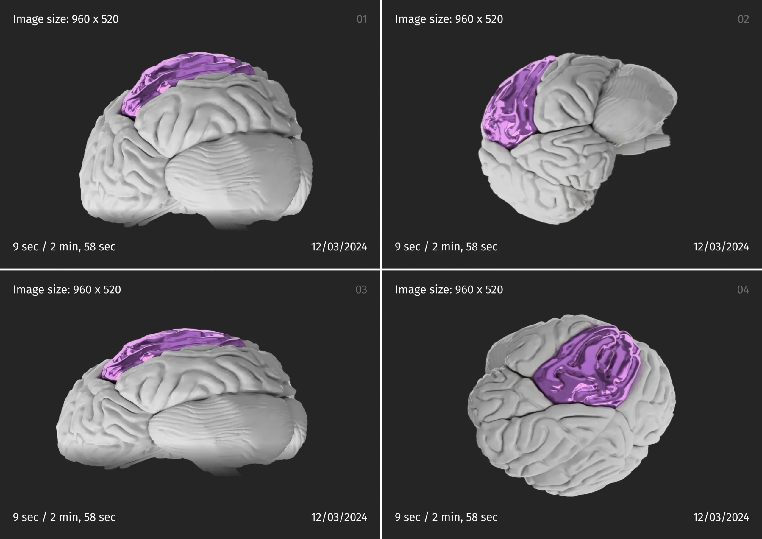
Our team developed an advanced ROI manager to highlight pathology. Doctors can easily identify and outline tumors in 3D reconstructions and measure lesion sizes to make informed surgical decisions.
Our developers set thresholds, pixel values, and previews for precise segmentation. This allows for detailed 3D customization in the form of reports with anatomical annotations and organ distance measurements, helping more accurate surgical planning. In addition, practitioners can export and share 3D images based on user access.
Results and business value
The 3D rendering platform allows professionals to monitor organs, evaluate tissue composition, assess fractures and thus diagnose diseases accurately. The platform generates detailed 3D medical imaging models and reports with anatomical annotations, as well as measures tumors, pathologies, and distances between organs for precise and effective surgical planning.
3X
faster pre-operational preparation
30%
more accurate diagnoses
Related cases
Need assistance with a software project?
Whether you're looking for expert developers or a full-service development solution, we're here to help. Get in touch!
What happens next?
An expert contacts you after thoroughly reviewing your requirements.
If necessary, we provide you with a Non-Disclosure Agreement (NDA) and initiate the Discovery phase, ensuring maximum confidentiality and alignment on project objectives.
We provide a project proposal, including estimates, scope analysis, CVs, and more.
Meet our experts!
Viktoryia Markevich
Relationship manager
Samuel Krendel
Head of partnerships

